Infrared ion spectroscopy, another dimension in mass spectrometry
Infrared ion spectroscopy, another dimension in mass spectrometry: application to the structural characterization of ETD generated peptide fragments
FELIX researchers have demonstrated the first use of infrared ion spectroscopy (IRIS) to characterize the structures of peptide fragments generated by electron transfer dissociation (ETD). They related previously predicted product ion structures of an often studied model tryptic peptide ([AlaAlaHisAlaArg+2H]^2) to experimental IR spectra of mass-selected ETD product ions to establish their molecular structure. Nature Communications publishes their results on June 9.
Comparing theory and experiment reveals that structural assignments based on computed thermodynamic considerations alone often do not correspond to the experimentally formed species. This highlights the potential for infrared ion spectroscopy using a tunable free electron laser (FELIX) to diagnostically identify product ion structures in tandem MS and, more specifically, the mechanisms associated with peptide fragmentation in electron attachment methods.
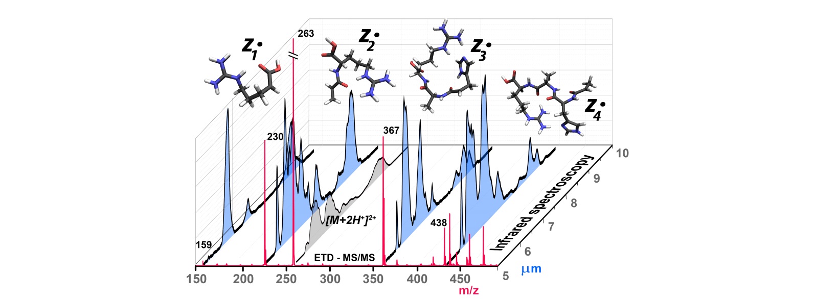
The IRIS method adds an extra dimension to a mass spectrum: for selected peaks in the MS, an IR spectrum is recorded. In combination with quantum-chemical calculations, the IR spectra reveal the molecular structure of the ions, which is undecipherable from the mass value alone. The technique is applied here to a series of product ions generated by electron transfer dissociation (ETD) of a short doubly protonated tryptic peptide (AlaAlaHisAlaArg + 2H+).
Furthermore, the results suggest that hydrogen atom transfer - necessary for radical migration - often does not occur after ETD without additional activation. This leads to the frequent observation of non-equilibrium product ions. Understanding intermolecular hydrogen atom migration is also of practical importance as it causes shifts in ETD fragment masses and makes sequence ion assignments more complicated.
Background
Developments in mass spectrometry drive many advances in the ‘omics’ sciences, including in particular proteomics. Identification of proteins, i.e. determining the sequence of amino acid residues constituting the protein, is routinely performed after mass-selective isolation of the protein in the gas phase environment of the MS, and characterization of the molecular weights of its fragments. Fragmentation is assumed to occur along the backbone and can be induced through a variety of methods. While collision-induced dissociation has been the standard for many years, alternative methods such as electron-induced dissociation are rapidly becoming more popular. Despite the routine application of these sequencing methods, the chemical mechanisms of the underlying dissociation reactions are often not accurately known. The mass of the molecular fragments alone does not provide molecular structure information from which reaction mechanisms could be inferred. Making use of the FELIX infrared free electron laser, IR spectra can be recorded for mass-selected molecular ions in an MS, revealing isomeric, tautomeric and conformeric structure information.
Reference:
Structural identification of electron transfer dissociation products in mass spectrometry using infrared ion spectroscopy
Jonathan Martens, Josipa Grzetic, Giel Berden, Jos Oomens, Nature Communications, DOI: 10.1038/ncomms11754
More information?
Jonathan Martens: j.martens@science.ru.nl
Jos Oomens: j.oomens@science.ru.nl